Content
-
1Anatomy of the reflex arch of the knee reflex
- 1.1Role and function in the body
- 1.2How to define it?
- 1.3Absence and reduction of the arc
- 1.4Hyporeflexia
- 1.5Hyperreflection
- 1.6Areflexia
- 1.7"Inspection of the knee reflex"
-
2Knee-jerk reflex and its meaning. Arc of the knee reflex
- 2.1How to cause a knee jerk?
- 2.2What if I need other methods?
- 2.3What are the violations in the work of the knee reflex?
- 2.4What is a reflex arc?
- 2.5What are the types of reflex arcs?
- 2.6How does the reflex arc of the knee reflex work?
- 2.7How does the impulse move at the knee reflex?
-
3Knee-jerk reflex
- 3.1How does this happen
- 3.2Scheme of the reflex arc of the knee reflex
- 3.3And how to check?
- 3.4Signs of a pathological reaction
- 3.5Hyperreflection
- 3.6Hypo- and areflexia
-
4Knee-jerk: components, arc diagram and its meaning
- 4.1The structure of the knee
- 4.2Types, function and meaning in medicine
- 4.3How to determine?
- 4.4Reduction and absence
-
5Reflex arc
- 5.1Types of reflexes
- 5.2What does the reflex arc consist of?
- 5.3Polysynaptic arches
- 5.4Arc of the blinking reflex
- 5.5Monosynaptic reflex arc
-
6What is the Achilles reflex: the scheme of the reflex arc, its description and verification
- 6.1What is an Achilles reflex
- 6.2Methods of research
- 6.3The reflex response from the Achilles tendon is reduced or completely lost in diseases:
- 6.4The reflex arc and the importance of its norm
- 6.5Diagnostic measures
- 6.6Symptoms of Violations in the Achilles Reflex
- 6.7Provocators of diseases
Anatomy of the reflex arch of the knee reflex
The knee-jerk reaction is the response of the body, which occurs with a slight stretching of the femoral muscle. The contraction of the muscles occurs as a result of a slight blow to the patella, under which is the tendon.
Under the external factor, the tendons stretch and activate the extensor muscle. This reflex is very important for diagnosing many diseases. But it is impossible to perform this procedure without a reflex arc.
The activity of the body depends on the reaction to the irritating receptors that come from the central nervous system. It is this structural basis of the reflex that is the reflex arc.
Reflex arc - the path of the transmitted signal from the receptor to the corresponding organ, which reacted to it. In another way it is called a nervous arc.
This name is explained by the fact that the knee reflex is performed due to nerve impulses that come through a certain pathway.
An arc is placed in the cells of the spinal cord, which, after excitation, can transmit an impulse to the muscles. The scheme with the designations of the reflex arc is not complicated, and it is possible to understand the functioning of the process with the help of a photo. There is a nerve arc of the following components:
- Links (central, efferent, afferent);
- Receptors;
- Effector (an organ that can change during the reflex).
Reflex arcs are of two types: simple and complex. Simple or monosynaptic reflex arcs consist of 2 neurons (efferent and afferent) and a synapse. Have the following features:
- Short duration of the reflex;
- Very close effector and receptor;
- The arc is two-neural;
- Muscles have a single muscle contraction;
- Neuron fibers of group A.
Complex or polysynaptic arcs contain in their composition three neurons (effector, receptor or pair of intercalary). Features of a complex nervous arc:
- The arc is three-neural;
- Nerve fibers of group B and C;
- The receptor and effector are not close together;
- Reduction of muscles according to the type of tetanus.
Role and function in the body
In simple words, the nerve arc is the path through which the impulse originates from the receptor to the organ or muscle.In accordance with this factor, the reflex arc is designed to transmit nerve impulses.
The pulse transfer scheme is based on the fact that the signal is transmitted from the receptor to the sensitive neurons. Further, the stimulating reaction is transmitted to the cells of the gray matter of the spinal cord.
As a result, motor cells contract, while the leg can jerk, or lift.
The impact acts as an external stimulus on the nervous system. Thanks to the connection between the spinal cord, the sensory system, the motor neurons, a process takes place. Visually present the description and understand the path of the flow of the nerve impulse will help the figure, where the nerve arc is depicted.
The receptors of the arc receive signals from the stimulus, and as a result of the feedback they are excited on them. The links perform the transfer of momentum to a specific organ. They are: central, efferent and afferent. The effector is the organ that responds to the action of the receptor.
According to these components of the arc, it will perform such functions:
- Transmits the signal to the muscle of the gastrocnemius;
- From neurons imparts an impulse to the motor muscles;
- Depending on the stimulus, the neuronal impulse transmits to the effector (organ);
- Affects the movement of the limb, muscle contraction of the leg.
How to define it?
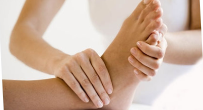
In order to correctly determine the presence of the knee reflex, the following steps must be performed:
- Place the patient in such a position on the chair so that he can freely throw his foot on the leg or that the limbs do not touch the floor.
- Then the doctor strikes the knee with a neurological hammer, triggering her response. These measures will help a specialist to determine the reflex arc of the knee.
Absence and reduction of the arc
The roots of gray matter can come into contact with other neurons. After that, they come into contact with the central neurons, forming the links of the conducting path. The reflex arc in this case may fail, as a result of the attachment of neurons to the spinal reflex.
Rapid excitations of the nervous system can be transmitted to the cerebral cortex and provoke new reflexes. As a result, irritation can return to the peripheral neuron, resulting in a complete absence of the knee reflex (areflexia).
Decrease reflex can through an intoxication of an organism, an infection, an epileptic seizure.
At rest, the knee arc is due to the pathology of the nervous system, the personal characteristics of the patient.
Pathological changes in the nervous system manifesting in the knee reflex may have a subsequent character: hyporeflexia, hyperreflexia and areflexia.
Hyporeflexia
- Irritant reaction in this pathology will decrease. A characteristic feature of this phenomenon is that the knee weakly reacts to the stimulus. There is a deviation due to a violation of the conductivity and integrity of the reflex arc during the transmission of the pulse along the neurons.
- Absence of a reflex may indicate a disease of the centers of the brain. Loss of body weight, infection lead to malnutrition of neurons and malfunctioning of cells. The reaction disappears after applying a tourniquet, anesthesia.
Hyperreflection
- The slightest influence on the limb leads to an increased knee reflex. It is often observed in the separation of the spinal cord. Since these structures block the impulses, in response to the stimulation.
- Occurs in persons of a neurotic type, with neuritis, plexitis, radiculitis. In addition, pathological movements, with a rapid contraction of the muscles of the stretched tendon, are an intensification of the reflex. Often they hit the foot and knee cap.
Areflexia
- It is a special type of pathology of the knee reflex, manifested as a result of the presence of a serious disease of the central nervous system. With such a pathological process, there is no irritant reaction to the imitative factor at all.
- Aereflexia occurs in the case of neuritis, poliomyelitis, polyneuritis, tabes. There is a lesion of a conducting neuron or motor neuron, sensitive fibers. Reduced reflex functions, coupled with damage to the nerve areas of the brain and spinal cord, the muscle reflexes are dying out.
"Inspection of the knee reflex"
How to conduct a neurologic examination by a specialist you can see in the next video.
A source: https://prospinu.com/anatomija/kolennyj-refleks.html
Knee-jerk reflex and its meaning. Arc of the knee reflex
Incorrect work of the knee reflex indicates serious disorders of the body. To diagnose the disease in the early stages, you should know what your reaction to the impact of the hammer under the knee tells you. Consider this in the article.
The reception of information from outside and its translation through the body: by muscles, organs, spinal cord and brain is ensured by the stable operation of nerves. The standard scheme for impulse transmission on the way is the brain.
In cases where an immediate reaction is required, the reflex passes through the spinal cord. This reaction occurs, for example, if you step on the needle, then the leg is sharply pulled back.
If the reflex were to go through the brain, there would definitely be a delay in the process, which is dangerous for the life of the organism.
So, the reflex is an instant response to an external stimulus, it is coordinated by the nervous system. And his way is called a reflex arc.
The stimulus signal is transmitted by afferent nerves to the efferent centers in the spinal cord. Then it is passed on to the muscles that contract.
Absence of reflexes is a symptom of a disease of the muscles, nervous system, brain, special emotional state.
Vital processes of the body also work reflexively, for example, the saliva supply when consuming food.
How to cause a knee jerk?
The origin of the knee reflex is due to the fact that during the impact of the medical malleus on the quadriceps tendon it contracts. This reduction causes the leg to straighten.
The impact must be applied exactly under the knee cap, because the tendon of the unbending quadriceps muscle is fixed at the beginning of the tibia.
It is not necessary to beat with force, the main thing is that the muscles are as relaxed as possible.
You can throw one foot to the other, then when the patellar reflex arises, it will jerk up.
What if I need other methods?
In the event that the traditional method does not work, there are several other techniques for manifesting the knee reflex:
- The person should be put on a chair so that the toes rest against the floor, and the legs bend at an angle of slightly more than 90 degrees. The blow must be applied from the top down over the protruding patella. As a result, the patella rises;
- The knee of the necessary leg should be placed on top of the second knee;
- You can apply a high seat so that your legs hang in a relaxed state;
- There is also a way when the patient is lowered onto the back with one knee in the other.
There are times when a patient physically can not sufficiently relax the examined limb.
Then the specialists apply the methods of knee-jerk disinhibition, for example, the techniques of Endrassic and Shvetsov.
Also, the patient should breathe deeply or aloud to solve simple mathematical examples.
What are the violations in the work of the knee reflex?
The muscles contract in a similar manner on the upper pair of limbs and in other parts of the body. But the value of the knee reflex is that its violation is considered an important symptom of abnormalities in the functioning of the brain and spinal cord.
The arc of the knee reflex is constant. Only in rare cases, a healthy person may not have a knee reflex, and, most likely, a child's disease has damaged his work.
In the presence of diseases, it may be absent or, on the contrary, excessively intensified. This is because the center of the knee reflex is located in the lumbar spinal cord, or rather in the II-IV segment.
.For some diseases, there are specific deviations in the manifestation of the knee reflex. For example, cerebral lesions cause a pendulum-like knee-jerk reflex. An intensified reflex may indicate a form of neurosis.
.On the contrary, the reduced form of the reflex is a sign of infection or intoxication of the body. A complete absence of the knee reflex indicates a significant lesion of the nervous system.
Also, the reflex may disappear in epileptics after a seizure, after using a tourniquet, during a deep anesthetic or after a heavy muscular load. Only an expert can make an accurate diagnosis.
What is a reflex arc?
The knee reflex is due to its reflex arc.
Like a significant disruption in the overall working process of the mechanism due to the presence of a damaged part, the human body can not function in the same way when something is not working properly.
The reflex arc is the path of the signal from the receptor that received it to the organ that reacts to it. It is also called a nerve arc.
This name is explained by the fact that the knee-jerk reflex occurs due to impulses in nerves that overcome a certain path. The reflex arc consists of chains of neurons that are formed from intercalary, receptor and effector neurons. They themselves and their processes create a path for transmission of irritation.
What are the types of reflex arcs?
The peripheral nervous system has two types of reflex arcs:
- Those that signal the internal organs;
- those that are related to skeletal muscles.
How does the reflex arc of the knee reflex work?
The arc of the knee reflex involves three parts of the back, from the second to the fourth. The fourth department is the most important in the process.
https://www.youtube.com/watc? = I-RB0Bwx58w
The reflex arc of the knee reflex has five components:
- Receptors. They receive the stimulus signal and are reactivated in response. These are the ends of axons or the corpuscles in the cells of the epithelium. Receptors are found everywhere in the human body, in organs, in the skin, of which sense organs are composed;
- Nerve fibers are sensitive, afferent or centripetal. It transmits the signal to the center. Neural bodies are located outside the central nervous system, namely near the brain and in the nerve nodes near the spinal cord.
- The nervous center is the place where the signal is transmitted from the afferent neurons to the efferent. The centers of efferent neurons are in the spinal cord.
- Nerve fibers are motor, centrifugal or efferent. As the name implies, the excitement on it goes from the central nervous system to a specific organ. The efferent fiber itself is an axon (or a long process) of a centrifugal neuron.
- The effector. An organ that exhibits a reaction to the irritation of a particular receptor. It is a muscle that contracts after processing the signal from the center, iron, which exudes juice due to nervous excitement, and more.
How does the impulse move at the knee reflex?
For a detailed study of the knee reflex, its stages should be studied. The passage of nerve impulses in the knee reflex occurs as follows:
- a hammer blow to the tendon under the knee causes this tendon to stretch, hence, a receptor potential arises in the corresponding receptors;
- In the neural long process, the potential for action arises. In the spinal cord, it is chemically transmitted to the motor neuron;
- The axon of the efferent neuron serves as a signal to the gastrocnemius muscle;
- because of the contraction of the muscle, the leg twitches.
Now you know how the reflex works, and for what purposes it is carried out by its diagnostics.
A source: http://.ru/article/227655/kolennyiy-refleks-cheloveka-i-ego-znachenie-duga-kolennogo-refleksa
Knee-jerk reflex
The knee-jerk reflex refers to the group of unconditioned reflexes and occurs when the quadriceps (quadriceps muscle of the thigh) is briefly stretched. Call this reflex with a light blow to the tendon area under the knee.
As a result of the impact, the tendon stretches, while the extensor muscle involuntarily contracts, and the leg is unbent at the knee joint. In other words, this is the response of the body to an external stimulus.
How does this happen
At the time of direct action on the tendon structure, sensitive receptors receive an impulse that is transmitted to the posterior horns of the spinal cord. The path through which the nervous signal passes is called a reflex arc.
A reflex arc, or a neural pulse path, is a concept that can refer to a somatic or autonomic nervous system.
The somatic department is responsible for the innervation of muscles, the autonomic nervous system supports the activity of internal organs, including blood vessels and various glands of external and internal secretion.
The neural path of the most simple muscle reflex is continuous and consists in the transmission of excitation between the two neurons. The reflex arcs of the autonomic nervous system are necessarily interrupted in the vegetative nodes (nervous ganglia).
The impulse path of the somatic nervous system is extremely simple, in the autonomic nervous system the signaling is somewhat complicated: there are additional nerve cells that convert the pulse from the sensitive receptor into a pulse for the effector organ.
The nervous impulse goes from the sensory to the motor neuron through the spinal cord
The pulse path consists of the following elements:
- A receptor that takes primary irritation or an external stimulus. It is a sensitive nerve ending that turns the factor of external action into a nerve impulse. There are many receptors in the human body that have different purposes. Those of them that are located close to the surface of the body are called exteroceptors.
- The sensitive nerve fiber is a long process of the neuron, which is located in the ganglion of the spinal cord.
- Effector neurons are the center that receives excitatory signals from sensitive nerve fibers. Simple reflexes have a center in the ganglions of the spinal cord, for complex reflexes the center is located in neurons of the brain.
- Motor nerve fibers directed towards the working part.
- The effector organ closes the arch and is represented by musculature, vessels or an internal organ.
Scheme of the reflex arc of the knee reflex
One of the examples of the neural path in the somatic nervous system is the tendon knee reflex, which is present in all people.
With neurological pathologies, the knee reflex may be reduced or, conversely, increased, so this neuropathologist tests this reaction in the first place.
The reflex arc of the knee reflex begins with the skin receptor, which reacts to the impact of the neurological mallet.
You can check the knee jerk even in a recumbent patient, for this the doctor simply lifts the limb, and operates the hammer on the hamstring
The resulting impulse is transmitted further to the neurons of the spinal cord, then passes into the appropriate center, to the effector neurons.
The long process of the effector neuron emerges from the spinal cord together with the motor nerve fibers that terminate in the muscles.
Thus, the impulse reaches the effector organ, that is, before the popliteal muscle, and the human foot abruptly moves upward: the knee reflex closes.
And how to check?
To find out whether the knee reflex is normal, a simple test is performed. The patient sits on a chair or a couch and puts one foot on the other. The main condition is the free position of the leg, which participates in the test: it should not rest on the floor.
The doctor strikes a slight blow to the neurological hammer in the area just below the knee, where the patella ends. The conclusion that the reflex persists follows from the immediate contraction of the quadriceps muscle of the thigh and the simultaneous rise of the tibia upwards.
The impact strength may be small, but the leg muscles should be necessarily relaxed.
If, for some reason, relaxation is not achieved and the patient tries to control his reactions, disinhibitory techniques are used.
For example, the subject is asked to carry out some kind of computational operation in his mind by adding or multiplying certain figures.
.It should be noted that there are several provisions in which the leg is maximally relaxed. Depending on the state of health or other specific conditions, the patient may lie during the test or sit without touching the floor.
.In the prone position, the tested leg is either held by the physician in weight, or the patient puts one foot on the knee of the other. The knee jerk is evaluated based on the amount of the angle of the shank deflection.
Signs of a pathological reaction
The normal knee reflex has an average degree of severity and is called normoflexia. If there are violations in the activity of the nervous system, then the passage of the neuronal impulse is hampered, which immediately reflects on the reflex reactions.
In healthy people, the reflex remains normal under any circumstances, except for patients with a tendency to neuroses. In medical practice, there are 3 types of abnormalities, when the knee-jerk:
- increased (hyperreflexia);
- decreased (hyporeflexia);
- absent (areflexion).
Any of the above deviations requires medical supervision and appropriate correction.
Hyperreflection
The increased knee-jerk reflex is characterized by a sharp deviation of the tibia by a significant angle and by almost complete extension of the leg in the joint. At the same time, a very weak effect on the popliteal ligament is enough to cause it.
Recommended revered: why pulls the leg under the knee from behind
Such a state can be observed with high excitability of the neurons of the anterior horns of the spinal cord due to low inhibitory control. That is, the anterior horns of cerebrospinal gray matter delay the signals coming from the brain in response to external influences.
Hyperreflexia is characteristic of spastic, or central, paresis and may be one of the symptoms of neuritis, plexitis, radiculitis, and also accompany various poisoning with poisonous substances.
With the strengthening of the knee reflex, there are so-called clones - spastic contractions of muscle structures due to stretching of the tendon.
The structure of the patella is such that if you grab its upper part and then release it sharply, the muscles of the quadriceps femoris will still twitch and contract for a while.
Hypo- and areflexia
Decrease in the response during a neurologic test indicates a violation of conduction in the femoral nerve, upper lumbar spinal nerves. In some cases, a low degree of knee reflex may indicate damage to the anterior horns of the spinal cord at the site of the exit of the nerve roots L3-L4.
The result of an epileptic fit is often the loss of reflexes, including the knee
Hyporeflexia is one of the signs of paresis of the lower extremities, a complete absence of a tendon reaction to irritation is observed due to a number of reasons:
- paralysis;
- physical exhaustion (cachexia of various etiologies);
- clamping of the thigh artery;
- an attack of epilepsy;
- after general anesthesia;
- dorsal dry.
Normal reflex reactions are an important diagnostic criterion for a neurologist.
Therefore, the knee reflex is checked first to draw conclusions about the state of the body.
In each specific case, an integrated approach is necessary, taking into account the individual characteristics of the patient and the results of the survey.
A source: http://MoyaSpina.ru/raznoe/kolennyy-refleks
Knee-jerk: components, arc diagram and its meaning
Everyone, even children, knows the procedure when, during a preventive medical examination, the neurologist beat on the knee, which in response rises sharply upward.
This amusing study is conducted in order to determine the knee jerk.
How important is this procedure, how correctly to conduct it, and how to record the result? - the answer to these questions can be found in this article.
The structure of the knee
Before talking about how the knee reflex arises, it is necessary to understand the structure of the knee joint and the mechanism of the appearance of a reflex in it. Speaking of the knee, most patients think about the joint itself, although the constituents of the knee are much larger, here they are:
- femur, and also tibia;
- meniscus;
- patella;
- condyles;
- muscle.
When there is a concept of how the knee joint is arranged, one can speak of the mechanism of the appearance of reflexes in it.
Reflex is a quick response of the body in response to an external stimulus. It is guided by the action of the reflex arc localized in the central nervous system of man. All reflexes are divided into two groups according to their origin:
- conditioned reflexes;
- reflexes of unconditional type.
Types, function and meaning in medicine
The patellar (knee) reflex is a reflex response of an unconditioned organism that occurs when the quadriceps femoris is briefly stretched.
Muscular contraction occurs as a result of an easy blow to the patella, under which the tendon is located.
With external stimulation, the tendon stretches, which activates the extensor muscle.
.This reflex is of great importance in neurology and diagnosis of many diseases.
.Thanks to an easy blow to the tendon, the doctor can immediately evaluate the operation of the femoral nerve and lumbar spine.
But this study is impossible without the reflex arc of the patellar reflex.
The reflex arc of the knee reflex, or, as it is also called, the neural pathway, is the pathway made by nerve impulses. There are two types of arcs:
- monosynaptic;
- two-neural.
The scheme of this reflex arc is quite simple, and you can understand it, knowing the main components, namely:
- receptors;
- links;
- effector.
Understand this process will help drawing, which shows the complete arc scheme and on which you can visually see the full path of the nerve impulse.
The receptor is the ends of the sensitive process of the axon, which receive signals from the stimulus and are excited in response to them.
The effector is the organ in which changes occur in response to the action of a particular receptor. In this case it is a muscle that shrinks in response to a hammer blow.
Based on the scheme of the reflex arc, the knee reflex can be divided into several stages:
- Stretching of the tendon, which occurs as a result of a hammer blow, receptor potential develops in the corresponding receptors.
- In the long process of the neuron, the potential of action is generated, which is chemically transferred to the body of the motor neuron.
- The signal to the gastrocnemius muscle comes from the axon of the efferent neuron.
- The muscle of the leg is shortened, while the characteristic movement of the leg is of a flexural nature.
How to determine?
To check the presence of the knee reflex correctly, you need to do the following:
- Offer the patient to sit on a chair, throwing one leg to the second, or sit on a high chair so that the lower limbs hang freely, without touching the floor.
- The doctor strikes a neurological hammer or an edge of his own hand on the area below the knee cap.
In neurology, another diagnostic technique is used to help determine the reflex arc of the knee reflex. When it is carried out, the patient is placed on a flat surface on his back.
The patient's lower limbs are bent at an angle of 100-1100, so that they are firmly resting their feet on the couch, and the blow is also applied to the tendon.
This method allows the doctor to see and evaluate the arc of the patellar reflex.
Reduction and absence
From the point of view of medicine, the presence of all reflexes testifies to the normal functioning of the nervous system, and consequently, of all organs.
If you yourself found yourself in the absence or weakness of the knee reflex, do not panic.
Entrust your health to a qualified neurologist who will conduct a study and make a conclusion.
There are several variants of deviation from the norm of the patellar reflex:
- hyporeflexia (spinal cord);
- hyperreflexia (diseases of the spinal cord);
- complete loss (organic lesions of the central nervous system, intoxications, infectious lesions, oncological cachexia).
Determine the presence of deviation from the norm and the degree of its severity will be able only by a highly qualified doctor who will necessarily appoint additional methods of investigation.
A source: http://SustavLife.ru/noga/kolennyi/reflektornaya-duga-kolennogo-refleksa.html
Reflex arc
The nervous activity of the human body is the transmission of impulses. One of the results of such programs is reflexes. In order for a certain reflex to be performed by the body, a connection must be established between the reception of the signal and the response to the stimulus.
Reflex arc
Types of reflexes
Reflex represents the reaction of a part of the body to the modification of the external or internal environment as a result of action on the receptors.
They can be found on the surface of the skin, giving rise to exteroreceptive reflexes, as well as on internal organs and vessels, which underlies the interorcesive or myostatic reflex.
Response reactions to stimuli are by nature conditional and unconditional. The second group includes reflexes, the arc of which is formed already at the time of birth. In the former it is created under the influence of external factors.
Types of reflexes
What does the reflex arc consist of?
The arc itself represents the entire path of the nerve impulse from the moment of contact of the person with the stimulus to the manifestation of the response. The reflex arc contains various types of neurons: receptor, effector and intercalary.
The reflex arc of the human body works like this:
- receptors perceive irritation. Most often, these receptors are the processes of nerve fibers of the centripetal type or neurons.
- The sensitive fiber translates excitation to the central nervous system. The structure of a sensitive neuron is such that its body is located outside the nervous system, they are ligated at the nodes along the spine and at the base of the brain.
- switching from a fiber of a sensitive type to a motor type occurs in the spinal cord. The brain is responsible for the formation of more complex reflexes.
- The motor fiber carries excitation to the reacting organ. This fiber is an element of the motor neuron.
The effector is actually the self-reacting organ, responds to irritation. Reflex response is contractile, motor or excretory.
The scheme of the origin of the reflex arc
Polysynaptic arches
The polysynaptic is a three-neuron arc, in which a nerve center is located between the receptor and the effector. Such an arc is clearly illustrated by the withdrawal of the hand in response to pain.
Polysynaptic arches have a special structure. Such a chain necessarily passes through the brain. Depending on the location of the neurons that process the signal, the following are distinguished:
- spinal cord;
- bulbar;
- mesencephalic;
- cortical.
If the reflex is processed in the upper parts of the central nervous system, then the neurons of the lower parts take part in its processing. The departments of the brainstem and the spinal cord also participate in the formation of high-level reflexes.
Whatever the reflex, if the continuity of the reflex arc is broken, then the reflex disappears. Most often, such a break occurs as a result of trauma or illness.
In complex reflexes, various organs are included in the chain links to react to the stimulus, which can change the behavior of the organism and its systems.
Arc of the blinking reflex
Arc of the blinking reflex
The structure of the arc of the blinking reflex is also interesting. This reflex, due to its complexity, makes it possible to study such an excitation motion along an arc, which it is difficult to investigate in other cases.
The reflex arc of this reflex begins with the activation of the exciting and inhibitory neurons simultaneously. Depending on the nature of the damage, various parts of the arc are activated.
To provoke the beginning of the blinking reflex, the trigeminal nerve can respond to a touch, the auditory response to a sharp sound, the visual response to a light drop or a visible danger.
.The reflex has an early and a late component. The late component is responsible for the formation of the response delay. As an experiment, touch the skin of the eyelid. The eye closes with lightning speed. When you touch the skin again, the reaction is slower.
.After brain processing of the received information, the acquired reflex is consciously inhibited. Thanks to such inhibition, for example, women very quickly learn to paint eyelids, overcoming the natural desire of the age to cover the cornea of the eye.
Other variants of the polysynaptic arches are also amenable to research, but they are often too complex and not very visible for study.
Whatever heights science has not reached, the basic reflexes for studying the human reaction remain blinking and knee-jerk reflexes.
The study and measurement of the rate of passage of the pulse in the trigeminal and facial nerves are the basis for assessing the state of the brain stem in various pathologies and pains.
Monosynaptic reflex arc
The arc, which consists of only two neurons, which is quite sufficient for the pulse, is called monosynaptic. A classic example of a monosynaptic arc is the knee jerk.
This is why a detailed diagram of the knee reflex arc is placed in all medical textbooks. A feature of the composition of such an arc is that it does not involve the brain. The knee-jerk reflex refers to the unconditioned muscular.
In man and other vertebrates such muscle reflexes are responsible for survival.
Pattern of the knee reflex
It is not surprising that it is the knee reflex that is checked by a neurologist as one of the indicators of the state of the somatic nervous system.
When the hammer strikes the tendon, the muscle stretches, after passing through the stimulation through the centripetal fiber to the spinal cord, the signal through the motor neuron into the centrifugal fiber.
In this experiment, the skin's receptors do not participate, however its result is very noticeable and the reaction strength is easily differentiated.
.The autonomic reflex arc breaks apart to form a synapse, whereas in the somatic system, the path that is overcome by the impulse from the receptor to the active skeletal muscle is not interrupted.
.A source: http://sustavam.ru/anatomiya/reflektornaya-duga/
What is the Achilles reflex: the scheme of the reflex arc, its description and verification
The name Achilles reflex got its name from the name of the legendary warrior Achilles, because his feet were very vulnerable.
The tendon that goes behind the leg on the shin to the beginning of the foot was named after the famous Greek with heel problems.
Physiologically, this unconditioned reflex, characterized by a profound manifestation, is formed in the child only at the age of 7 days.
The neonatologist fixes its formation by means of the influence of the percussion malleus on the Achilles ligaments. Reacts to the mechanical action of the triceps muscles of the lower leg, it contracts, behind this the feet are reflexively bent.
What is an Achilles reflex
How the Achilles tendon looks like
Under the definition of "Achilles reflex" in medicine is understood reflex reduction of muscles on calves of legs - tricipitis surae, occurring simultaneously with the plantar response to a hammer blow at the points of the heel tendons. This reflex has a deep physiological nature. The scheme of the Achilles reflex includes a response to the external stimulus of muscle tissue and tendons from the knee ligaments, through the calf muscles and tendons on the outside of the heels.
A simple reflex arc, the pattern of which closes in the tissues of the central nervous system, improves the adaptation of the person in the surrounding environment - involuntarily withdraw the hand from the hot iron, adjust the size of the pupils when changing illuminance.
The group of constant physiological responses of the body to external stimuli present in healthy people throughout life includes the Achilles reflex, a description which today is not difficult, as he is thoroughly studied by doctors, is well represented in the educational and methodical literature for medical training institutions.
The accepted method for determining the presence of a normal reflex - the patient must kneel so that the feet without tension hang from the chair. To completely relax the legs, the patient needs to tightly squeeze the back of the chair with his hands.
In this position, the Achilles reflex is checked. Examining the children, the doctor takes the palm of both feet with the palm of the hand, the child lies on the tummy, the legs bend at the knees.
The doctor fixes the legs in the middle of the bending, accurately and accurately taps the percussion hammer at the critical points of the Achilles tendons.
Methods of research
Checking Achilles Reflexes
There is a method of testing achilles reflexes, when a person lies on his back.
The neurologist with the help of the left hand fixes the right foot of the patient, pressing causes bending of the leg in the knee and hip joint, folding of the foot in the rear with simultaneous relaxation, after which it makes a survey and strikes the percussion hammer on sensitive points.
The reflex response from the Achilles tendon is reduced or completely lost in diseases:
- dorsal is the third stage of syphilis;
- polyneuritis - a massive lesion of nerve tissues and endings;
- sciatic nerve neuropathy;
- radiculitis;
- myelodysplasia;
- radicular lesion of the spinal cord;
- diseases of peripheral NA.
An increase in the reflex response, up to the clone of the foot, is a sign of the defeat of pyramidal pathways.
The reflex arc and the importance of its norm
Reflex arc of knee joint
The arc of the reflex response to the stimulus involves the reaction of the tissues of the tibial nerve, connected by nerve fibers to the spinal cord in segments S1-S2. The reflex arises as a result of the movement of the three-headed muscles of the shins.
The doctor needs to see the power of the reflex reaction. With repeated exposure to the percussion hammer, the reaction of the response varies, but remains within the regulatory framework.
If the reflex response decreases or rises, the doctor considers the situation of dysfunctions in bone structures.
The nervous reflex arc is the path that the impulse passes through. It:
- receptor that takes the action of the stimulus;
- Afferent centripetal link - processes of neurons that transmit impulse in the central nervous system;
- the central link;
- efferent link;
- effector - an organ that executes a command of a nerve signal.
The simplest example of a reflex arc involves the participation of two neurons - sensory and motor.
The task of the sensory neuron is to transmit an impulse to the brain, where the information obtained is processed by the corresponding nervous centers, and then sent downward to the spinal motor motoneurons, giving a change in the movements of the tested bodies. The arc of the pulse path can be closed, providing rhythmic movements. A weak impulse is an indicator of the disease in the examined sections of the central nervous system.
Some diseases cause a decrease in the reflex response. It:
- the spinal cord shows the processes of degradation;
- atrophic vertebral muscles;
- VSD;
- defeat of motor neurons.
With such serious pathologies, the fibers and endings of nerve tissues in the brain of the back and head are affected.
There is no achilles reflex if the patient being examined is not sick, but there is still no reaction. In this situation, the doctor reads the medical history, where he finds a description of the corresponding complaints of the patient.
A doctor's suspicion always causes the absence or significant weakening of the Achilles reflex, since this is most likely a sign of diseases that begin to develop in the bone structures of the spine, in the dorsal brain.
.Given that the reflex arc of the Achilles reflex extends through the lumbar and tibial spine and affects the vertebrae S5 lumbar and S1 sacral divisions, the physician assumes the corresponding deviations in the state of health caused by even old injuries.
.The most dangerous diseases:
- Radiculitis.
- Lumbosacral osteochondrosis.
- Intervertebral hernia.
With such diseases, there is a jamming of the nerve channels, the signal flow through the receptors is disturbed. Develops areflexia - the decline of reflexes from muscle tissue.
Areflexia is caused by special diseases that evenly affect all parts of the nervous connections of the spinal cord and brain.
The absence of the Achilles reflex is considered a pathological phenomenon.
Diagnostic measures
Diagnosis of Achilles reflexes
Importance of diagnostics of achilles reflexes consists in an opportunity to reveal illnesses of a backbone at an early stage.
At the same time, the absence of changes in the reaction does not affect the way people live.
Nevertheless, it is important to detect a failure in the functions of the spine in time, which is facilitated by the definition of a decrease or increase in the muscular reaction indicating the onset of the disease.
When such a diagnosis as an achilles reflex is assumed, the examination scheme is made for each patient individually, and depends on the symptoms that appear.
Therefore, the doctor prescribes a more accurate instrumental examination, which is most correctly performed in a stationary setting.
A X-ray of the lumbosacral section in several projections is mandatory, a study of cerebrospinal fluid, a study of blood on the RW to find the causes of the disease, for its effective treatment.
Symptoms of Violations in the Achilles Reflex
Violation of the functions of the injured limbs is reflected in the corresponding symptomatology, which disturbs the person when walking, doing normal movements, working with weights.
The main symptoms are:
- pain in the sacral region;
- numbness of the legs;
- constant freezing of the feet;
- increased excitability of the nerves of the back;
- high strength of muscle reaction;
- disturbance of the reflex arc up to paralysis of the nerves;
- lack of support in the forefoot;
- decreased muscle tone;
- atrophy of the triceps;
- lameness when walking.
The treatment is aimed at restoring the spent functions, restoring intervertebral connections.
In the course of therapeutic and even surgical treatment of Achilles, the reflex may not recover, completely absent, but this fact will simply remain in memory of the transferred disease, without affecting the quality life.
Types of diseases of the Achilles tendon:
- Tendonitis.
- Peritendinitis.
- Enthesopathy.
- Tendopathy.
All this - inflammatory processes, with which the treatment scheme is associated, which has the purpose to remove inflammatory processes and restore the motor functions of the legs. Women are immediately banned high heels, men are imposed restrictions in sports training related to the load on their feet.
Provocators of diseases
The main factors provoking the disease are:
- decrease in elasticity due to age-related changes;
- motor stresses leading to damage to the tendons;
- some forms of flatfoot, accompanied by hyperpredation, at which the sole foot wrapping is observed inside when walking;
- uncomfortable shoes, resulting in an incorrect load distribution.
A source: https://NogoStop.ru/golenostop/suxozhiliya/axillov-refleks.html