Pulmonary edema is the cause of the painful death of many patients. It occurs most often as a complication in the violation of the regulation of fluid volumes that must circulate in the lungs.
At this point, there is an active inflow of fluid from the capillaries into the pulmonary alveoli, which overflow with exudate and lose the ability to function and take oxygen. The man stops breathing.
What is this
Thisan acute pathological condition that threatens life, which requires extremely urgent care, immediate hospitalization. The main characteristics of the disease are characterized by acute air deficiency, severe suffocation and death of the patient in the absence of resuscitation.
At this point, active filling of capillaries with blood and rapid passage of fluid through the walls of capillaries into the alveoli occurs, where so much is collected that it greatly hinders the flow of oxygen. In respiratory organs, gas exchange is disrupted, tissue cells undergo acute oxygen deficiency ( hypoxia), the person suffocates. Quite often choking happens at night during sleep.
Classification, from which the
happens. The causes and types of pathology are closely related, divided into two basic groups.
| Hydrostatic( cardiogenic or cardiac) pulmonary edema | |
Occurs during diseases that are characterized by increased pressure( hydrostatic) inside the capillaries and further penetration of plasma into the pulmonary alveoli. The reasons for this form are:
| |
| Non-cardiogenic pulmonary edema, which includes: | |
| Iatrogenic | There is:
|
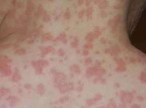
| Allergic, toxic( membranous) | Provoked by the action of poisons, toxins that break the permeability of the walls of the alveoli, when instead of air in them penetrates the liquid, filling almost the entire volume. Causes of toxic pulmonary edema in humans:
Often passes without any characteristic signs. The picture becomes clear only when carrying out radiography. |
| Infectious | Develops:
|
| Aspiration | Occurs when a foreign body enters the lungs, the contents of the stomach. |
| Traumatic | Happens with penetrating chest injuries. |
| Cancerous | Occurs because of a malfunction of the pulmonary lymphatic system with difficulty in lymph drainage. |
| Neurogenic | Main causes:
|
In these conditions, the alveoli become very thin, their permeability increases, the integrity of the is compromised, the risk of filling them with a liquid is increased.
Risk Groups Since the pathogenesis of pathology is closely related to concomitant internal diseases of , patients with diseases or factors that cause such a health and life-threatening condition are at risk.
The risk group includes patients suffering from:

- disorders of the vascular system, heart;
- with cardiac muscle damage in hypertension;
- congenital heart disease, respiratory system;
- with complex craniocerebral trauma, cerebral hemorrhages of different origin;
- meningitis, encephalitis;
- by cancerous and benign neoplasms in the brain tissues.
- pneumonia, emphysema, bronchial asthma;
- with deep vein thrombosis and increased blood viscosity;there is a high probability of detachment of a floating( floating) clot from the artery wall with penetration into the pulmonary artery, which is blocked by a thrombus, which causes thromboembolism.
In climbers such a dangerous condition happens with a rapid rise to a high altitude without holding a pause on intermediate high-level tiers.
Symptoms: as manifested and developed by stages of
Classification and symptoms are related to the degree of severity of the disease.
| Severity | Severity of symptoms |
| 1 - at the developmental boundary | Revealed:
|
| 2 - medium | Observed:
|
| 3 - severe | Clearly expressed symptoms:
|
| 4th grade - critical | Classical manifestation of critical condition:
|
First urgent first aid: what to do if you have an
Before the ambulance arrives, relatives, friends, colleagues of should not lose a minute of time .To facilitate the patient's condition, do the following:

- Help a person to sit or half-raise with their feet
- If possible, diuretics are treated( give diuretics - lasix, furosemide) - this removes excess fluid from the tissues, however, uses low doses of drugs at low pressure.
- Organize the possibility of maximum access to oxygen in the room.
- Foam suction is carried out and, at skill - perform oxygen inhalations through a solution of ethyl alcohol( 96% of the pair to adults, 30% of alcohol pairs to children).Prepare a hot foot bath.
- At skill - apply imposing on finitenesses of plaits , not too tightly pressing veins in the top third of a femur. Leave the tourniquets for more than 20 minutes, while the pulse should not be interrupted below the places of application. This reduces the flow of blood to the right atrium and prevents tension in the arteries. When the strands are removed, this is done cautiously, slowly loosening them.
- Continuously monitor how the patient breathes, at the pulse rate.
- With pain, analgesics are given, if there is - promedol.
- With high blood pressure, use benzohexonium, pentamine, which promote the flow of blood from the alveoli, nitroglycerin, dilating blood vessels( with regular measurement of pressure).
- At normal - small doses of nitroglycerin under the control of pressure indicators.
- If the pressure is below 100/50 - dobutamine, dopmin, increasing the function of contraction of the myocardium.
Than dangerous, forecast
Pulmonary edema is a direct threat to life .Without the extremely urgent measures that the patient's relatives should take, without the subsequent urgent active therapy in the hospital, pulmonary edema is the cause of death in 100% of cases. A person is waiting for suffocation, coma, death.
Preventive measures
In order to prevent a health and life threat, the following measures are presumed, meaning elimination of the factors contributing to this condition:

- In cases of heart diseases( angina, chronic insufficiency), funds are taken for their treatment and, at the same time, hypertensive disease.
- For repeated swelling of the respiratory system, the procedure of isolated ultrafiltration of blood is used.
- Operative precise diagnostics.
- Timely adequate treatment for asthma, atherosclerosis, other internal disorders that can cause such a pulmonary pathology.
- Isolate the patient from contact with any kind of toxins.
- Normal( not excessive) physical, as well as respiratory load.
Complications of
Even if in the hospital they quickly and successfully managed to prevent the suffocation and death of a person, the therapy continues. After such a critical condition for the entire body, patients often develop serious complications of , most often in the form of recurring pneumonia, which is difficult to treat.
Prolonged oxygen starvation has a negative effect on almost all organs. The most serious consequences are cerebral circulation disorders, heart failure, cardiosclerosis, ischemic lesions of organs. These diseases carry a constant threat to life and do not do without intensive drug therapy.
The greatest danger of this pathology is its rapidity and panic state of , into which the patient and the people surrounding him are emptying.
Knowledge of the basic signs of the development of pulmonary edema, the causes, diseases and factors that can provoke it, as well as emergency measures before the arrival of the ambulance can lead to a favorable outcome and the absence of consequences even with such a serious threat to life.